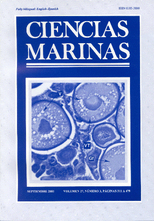
Histology of the oocytes of Lutjanus peru (Nichols and Murphy, 1922) (Pisces: Lutjanidae)
Main Article Content
Abstract
The distinctive characteristics found in each of the development stages of the oocyte in the ovary of Lutjanus peru (Pacific red snapper) are described. The histological technique was used to identify the development stages of the oocytes and the averages of the diameters of the oocytes were obtained for these stages. Stage I, chromatin nucleolus oocyte: average diameter of 52.89 µm. Stage II, perinucleolus oocyte: average diameter of 117.78 µm. Stage III, yolk vesicle oocyte: average diameter of 166.18 µm. Stage IV, primary vitellogenesis oocyte: average diameter of 221.73 µm. Stage V, secondary vitellogenesis oocyte: average diameter of 333.7 µm. Stage VI, tertiary vitellogenesis oocyte: average diameter of 340.17 µm. Stage VII, mature oocyte: average diameter of 340.17 µm. With the identification of these stages, the observation of the process of the oogenesis in L. peru has been completed.
Downloads
Article Details
This is an open access article distributed under a Creative Commons Attribution 4.0 License, which allows you to share and adapt the work, as long as you give appropriate credit to the original author(s) and the source, provide a link to the Creative Commons license, and indicate if changes were made. Figures, tables and other elements in the article are included in the article’s CC BY 4.0 license, unless otherwise indicated. The journal title is protected by copyrights and not subject to this license. Full license deed can be viewed here.

