
Patrones de fluorescencia y diversidad de hidrozoos en Bocas del Toro, Panamá
Barra lateral del artículo
Contenido principal del artículo
Resumen
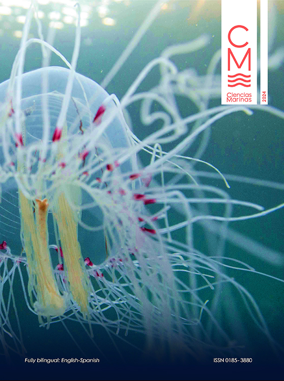
Muchos metazoos contienen moléculas capaces de fluorescencia, absorción y reemisión de luz. Dado que la distribución anatómica, o el patrón, de estas moléculas es variable entre taxones, los patrones de fluorescencia pueden servir como una poderosa herramienta diagnóstica en taxonomía y ecología. Sin embargo, los patrones de fluorescencia específicos de especie entre invertebrados marinos están poco comprendidos. Aquí, mostramos que los hidrozoos tienen diversos patrones de fluorescencia, que pueden ser el resultado de moléculas producidas intrínsecamente u obtenidas de fuentes dietéticas. Estudiamos hidrozoos, incluidos sifonóforos, hidromedusas e hidroides, en 5 órdenes de ambientes marinos pelágicos y bentónicos en Bocas del Toro, Panamá. Nuestros hallazgos muestran que los patrones de fluorescencia son muy prevalentes y pueden variar entre especies de hidrozoos y a lo largo del desarrollo. La mayoría de los taxa de hidrozoos examinados exhibieron alguna forma de fluorescencia, con variación observada entre las etapas de vida y las partes del cuerpo. Se documentó fluorescencia en el 88% de las hidromedusas (Leptothecata, Anthoathecata, Limnomedusae y Narcomedusae), el 50% de los hidroides y el 75% de los sifonóforos que se observaron en este estudio. Nuestros resultados ilustran cómo los patrones de fluorescencia pueden servir como una herramienta diagnóstica útil para explorar la biodiversidad marina, resaltando la necesidad de una mayor documentación de los patrones de fluorescencia en la diversidad marina y de investigar las moléculas que subyacen a este fenómeno.
Descargas
Detalles del artículo

Esta obra está bajo una licencia internacional Creative Commons Atribución 4.0.
Este es un artículo de acceso abierto distribuido bajo una licencia Creative Commons Attribution 4.0, que le permite compartir y adaptar el trabajo, siempre que dé el crédito apropiado al autor o autores originales y la fuente, proporcione un enlace a Creative Commons licencia, e indicar si se realizaron cambios. Las figuras, tablas y otros elementos del artículo están incluidos en la licencia CC BY 4.0 del artículo, a menos que se indique lo contrario. El título de la revista está protegido por derechos de autor y no está sujeto a esta licencia. La escritura de licencia completa se puede ver aquí.
Métrica
Citas
Aglyamova GV, Hunt ME, Modi CK, Matz MV. 2011. Multi-colored homologs of the green fluorescent protein from hydromedusa Obelia sp. Photochem Photobiol Sci. 10:1303-1309. https://doi.org/10.1039/c1pp05068k
Alieva NO, Konzen KA, Field SF, Meleshkevitch EA, Hunt ME, Beltran-Ramirez V, Miller DJ, Wiedenmann J, Salih A, Matz MV. 2008. Diversity and evolution of coral fluorescent proteins. PLOS ONE. 3(7):e2680. https://doi.org/10.1371/journal.pone.0002680
Alvariño A. 1974. Distribution of siphonophores in the regions adjacent to the Suez and Panama canals. Fish Bull. 72.
Bentlage B, Collins AG. 2021. Tackling the phylogenetic conundrum of Hydroidolina (Cnidaria: Medusozoa: Hydrozoa) by assessing competing tree topologies with targeted high-throughput sequencing. PeerJ 9:e12104 https://doi.org/10.7717/peerj.12104
Boero F, Bouillon J, Gravili C, Miglietta MP, Parsons T, Piraino S. 2008. Gelatinous plankton: irregularities rule the world (sometimes). Mar Ecol Prog Ser. 356:299–310. https://doi.org/10.3354/meps07368
Bomati EK, Manning G, Deheyn DD. 2009. Amphioxus encodes the largest known family of green fluorescent proteins, which have diversified into distinct functional classes. BMC Evol Biol. 9:77. https://doi.org/10.1186/1471-2148-9-77
Calder DR. 2019. On a collection of hydroids (Cnidaria, Hydrozoa) from the southwest coast of Florida, USA. Zootaxa. 4689(1):1-141. https://10.11646/zootaxa.4689.1.1
Chudakov DM, Matz MV, Lukyanov S, Lukyanov KA. 2010. Fluorescent proteins and their applications in imaging living cells and tissues. Physiol Rev. 90:1103-1163. https://doi.org/10.1152/physrev.00038.2009
Cornelius PFS. 1995. North-West European Thecate Hydroids and Their Medusae. Synopses of the British Fauna 50: Linnean Society of London. 347 p.
Cunha AF, Maronna MM, Marques AC. 2016. Variability on microevolutionary and macroevolutionary scales: a review on patterns of morphological variation in Cnidaria Medusozoa. Organisms Diversity & Evolution. 16:431-442. https://doi.org/10.1007/s13127-016-0276-4
De Campos CJA, Migotto AE, Pinheiro U, Marques AC. 2012. Sponges as substrata and early life history of the tubulariid Zyzzyzus warreni (Cnidaria: Hydrozoa) in the São Sebastião Channel, Brazil. Mar Biol Res. 8:573-583. https://doi.org/10.1080/17451000.2011.638641
Fourrage C, Swann K, Gonzalez-Garcia JR, Campbell AK, Houliston E. 2014. An endogenous green fluorescent protein-photoprotein pair in Clytia hemisphaerica eggs shows co-targeting to mitochondria and efficient bioluminescence energy transfer. Open Biology. 4:130206. https://doi.org/10.1098/rsob.130206
Francis WR, Christianson LM, Powers ML, Schnitzler CE, Haddock SHD. 2016. Non-excitable fluorescent protein orthologs found in ctenophores. BMC Evol Biol. 16:167. https://doi.org/10.1186/s12862-016-0738-5
Global Biodiversity Information Facility [GBIF]. 2023. GBIF Home Page: GBIF; [accessed 2023 Sept 15]. https://www.gbif.org.
Gravili C. 2016. Zoogeography of Hydrozoa: Past, Present and a Look to the Future. In: Goffredo S, Dubinsky Z (eds.), The Cnidaria, Past, Present and Future: The World of Medusa and Her Sisters. Cham (Switzerland): Springer International Publishing. p. 95-107. https://doi.org/10.1007/978-3-319-31305-4_7
Gravili C, Boero F, Alifano P, Stabili L. 2012. Association between luminous bacteria and Hydrozoa in the northern Ionian Sea. J Mar Biol Assoc UK. 92:1317-1324. https://doi.org/10.1017/S0025315411001408
Gruber DF, Kao H-T, Janoschka S, Tsai J, Pieribone VA. 2008. Patterns of fluorescent protein expression in Scleractinian corals. Biol Bull. 215:143-154. https://doi.org/10.2307/25470695
Haddock SHD, Dunn CW. 2015. Fluorescent proteins function as a prey attractant: experimental evidence from the hydromedusa Olindias formosus and other marine organisms. Biology Open. 4:1094-1104. https://doi.org/10.1242/bio.012138
Haddock SHD, Dunn CW, Pugh PR, Schnitzler CE. 2005. Bioluminescent and red-fluorescent lures in a deep-sea siphonophore. Science. 309:263. https://doi.org/10.1126/science.1110441
Hunt ME, Modi CK, Aglyamova GV, Ravikant DVS, Meyer E, Matz MV. 2012. Multi-domain GFP-like proteins from two species of marine hydrozoans. Photochem Photobiol Sci. 11:637-644. https://doi.org/10.1039/c1pp05238a
[Invertebase STRI] Smithsonian Tropical Research Institute. 2023. STRI Symbiota Portal: STRI; [accessed 2018 Aug 01]. https://invertebase.org/stri/.
Kramp PL. 1959. The hydromedusae of the Atlantic Ocean and adjacent waters. Dana Report. 46:1-283.
Kubota. 2010. Various distribution patterns of green fluorescence in small hydromedusae. Kuroshio Biosphere. 6:11-14.
Kubota, Shin, Gravili, Cinzia. 2011. Rare distribution of green fluorescent protein (GFP) in hydroids from Porto Cesareo, Lecce, Italy, with reference to biological meaning of this rarity. Biogeography. 13:9-11.
Kubota S, Pagliara P, Gravili C. 2008. Fluorescence distribution pattern allows to distinguish two species of Eugymnanthea (Leptomedusae: Eirenidae). J Mar Biol Assoc UK. 88:1743-1746. https://doi.org/10.1017/S0025315408002580
Labas YA, Gurskaya NG, Yanushevich YG, Fradkov AF, Lukyanov KA, Lukyanov SA, Matz MV. 2002. Diversity and evolution of the green fluorescent protein family. PNAS. 99:4256-4261. https://doi.org/10.1073/pnas.062552299
Lakowicz JR. 2006. Instrumentation for Fluorescence Spectroscopy. In: Lakowicz JR (ed.), Principles of Fluorescence Spectroscopy. Boston (USA): Springer US. p. 27-61. https://doi.org/10.1007/978-0-387-46312-4_2
Lamb JY, Davis MP. 2020. Salamanders and other amphibians are aglow with biofluorescence. Sci Rep. 10:2821. https://doi.org/10.1038/s41598-020-59528-9
Lim MLM, Land MF, Li D. 2007. Sex-specific UV and fluorescence signals in jumping spiders. Science. 315:481. https://doi.org/10.1126/science.1134254
Macel M-L, Ristoratore F, Locascio A, Spagnuolo A, Sordino P, D’Aniello S. 2020. Sea as a color palette: the ecology and evolution of fluorescence. Zoological Letters. 6:9. https://doi.org/10.1186/s40851-020-00161-9
Maggioni D, Montano S, Seveso D, Galli P. 2016. Molecular evidence for cryptic species in Pteroclava krempfi (Hydrozoa, Cladocorynidae) living in association with alcyonaceans. Systematics Biodivers. 14:484-493. https://doi.org/10.1080/14772000.2016.1170735
Maggioni D, Saponari L, Seveso D, Galli P, Schiavo A, Ostrovsky AN, Montano S. 2020. Green fluorescence patterns in closely related symbiotic species of Zanclea (Hydrozoa, Capitata). Diversity. 12:78. https://doi.org/10.3390/d12020078
Marshall J, Johnsen S. 2017. Fluorescence as a means of colour signal enhancement. Philos Trans R Soc B Biol Sci. 372:20160335. https://doi.org/10.1098/rstb.2016.0335
Masuda H, Takenaka Y, Yamaguchi A, Nishikawa S, Mizuno H. 2006. A novel yellowish-green fluorescent protein from the marine copepod, Chiridius poppei, and its use as a reporter protein in HeLa cells. Gene. 372:18-25. https://doi.org/10.1016/j.gene.2005.11.031
Matz MV, Fradkov AF, Labas YA, Savitsky AP, Zaraisky AG, Markelov ML, Lukyanov SA. 1999. Fluorescent proteins from nonbioluminescent Anthozoa species. Nat Biotechnol. 17:969-973. https://doi.org/10.1038/13657
Mazel CH, Cronin TW, Caldwell RL, Marshall NJ. 2004. Fluorescent enhancement of signaling in a mantis shrimp. Science. 303:51. https://doi.org/10.1126/science.1089803
Mazel CH, Lesser MP, Gorbunov MY, Barry TM, Farrell JH, Wyman KD, Falkowski PG. 2003. Green-fluorescent proteins in Caribbean corals. Limnol Oceanogr. 48:402-411. https://doi.org/10.4319/lo.2003.48.1_part_2.0402
Miglietta MP, Piraino S, Pruski S, Alpizar-Gonzalez M, Castellanos-Iglesias S, Jerónimo-Aguilar S, Lawley JW, Maggioni D, Martell L, Matsumoto Y, et al. 2018. An integrative identification guide to the Hydrozoa (Cnidaria) of Bocas del Toro, Panama. Neotropical Biodiversity. 4:103-113. https://doi.org/10.1080/23766808.2018.1488656
Montenegro J, Collins AG, Hopcroft RR, Questel JM, Thuesen EV, Bachtel TS, Bergman LA, Sangekar MN, Drazen JC, Lindsay DJ. 2023. Heterogeneity in diagnostic characters across ecoregions: A case study with Botrynema (Hydrozoa: Trachylina: Halicreatidae). Front Mar Sci. 9. https://doi.org/10.3389/fmars.2022.1101699
Oliveira OMP, Miranda TP, Araujo EM, Ayón P, Cedeño-Posso CM, Cepeda-Mercado AA, Córdova P, Cunha AF, Genzano GN, Haddad MA, . 2016. Census of Cnidaria (Medusozoa) and Ctenophora from South American marine waters. Zootaxa. 4194(1):1-256. https://doi.org/10.11646/zootaxa.4194.1.1
Oswald F, Schmitt F, Leutenegger A, Ivanchenko S, D’Angelo C, Salih A, Maslakova S, Bulina M, Schirmbeck R, Nienhaus GU, et al. 2007. Contributions of host and symbiont pigments to the coloration of reef corals. FEBS J. 274:1102-1109. https://doi.org/10.1111/j.1742-4658.2007.05661.x
Pakhomov AA, Martynov VI. 2011. Probing the structural determinants of yellow fluorescence of a protein from Phialidium sp. Biochem Biophys Res Commun. 407:230-235. https://doi.org/10.1016/j.bbrc.2011.03.004
Patry W, Knowles T, Christianson L, Howard M. 2014. The hydroid and early medusa stage of Olindias formosus (Cnidaria, Hydrozoa, Limnomedusae). J Mar Biol Assoc UK. 94:1409-1415. https://doi.org/10.1017/s0025315414000691
Petushkov VN, Gibson BG, Lee J. 1995. The yellow bioluminescence bacterium, Vibrio fischeri Y1, contains a bioluminescence active riboflavin protein in addition to the yellow fluorescence FMN protein. Biochem Biophys Res Commun. 211:774-779. https://doi.org/10.1006/bbrc.1995.1880
Pugh PR, Haddock SHD. 2010. Three new species of resomiid siphonophore (Siphonophora: Physonectae). J Mar Biol Assoc U K. 90:1119-1143. https://doi.org/10.1017/S0025315409990543
Rodriguez CS, Pujol MG, Mianzan HW, Genzano GN. 2014. First record of the invasive stinging medusa Gonionemus vertens in the southern hemisphere (Mar del Plata, Argentina). Lat Am J Aquat Res. 42:653-657. https://doi.org/10.3856/vol42-issue3-fulltext-23
Schnitzler CE, Keenan RJ, McCord R, Matysik A, Christianson LM, Haddock SHD. 2008. Spectral diversity of fluorescent proteins from the anthozoan Corynactis californica. Mar Biotechnol. 10:328-342. https://doi.org/10.1007/s10126-007-9072-7
Schuchert P. 2012. North-West European athecate hydroids and their medusae: keys and notes for the identification of the species: Field Studies Council. Report No. 59.
Schuchert P, Collins R. 2021. Hydromedusae observed during night dives in the Gulf Stream. Revue suisse de Zoologie. 128:237-356. https://doi.org/10.35929/RSZ.0049
Shagin DA, Barsova EV, Yanushevich YG, Fradkov AF, Lukyanov KA, Labas YA, Semenova TN, Ugalde JA, Meyers A, Nunez JM, et al. 2004. GFP-like proteins as ubiquitous metazoan superfamily: evolution of functional features and structural complexity. Mol Biol Evol. 21:841-850. https://doi.org/10.1093/molbev/msh079
Shimomura O. 2005. The discovery of aequorin and green fluorescent protein. J Microsc. 217:1-15. https://doi.org/10.1111/j.0022-2720.2005.01441.x
Steyaert M, Mogg A, Dunn N, Dowell R, Head CEI. 2022. Observations of coral and cryptobenthic sponge fluorescence and recruitment on autonomous reef monitoring structures (ARMS). Coral Reefs. 41:877-883. https://doi.org/10.1007/s00338-022-02283-2
Suggett DJ, Borowitzka MA, Prášil O. 2010. Chlorophyll a Fluorescence in Aquatic Sciences: Methods and Applications: Springer Science & Business Media. 332 p. https://doi.org/10.1007/978-90-481-9268-7
Yang H, Xiao X, Zhao XS, Hu L, Xue XF, Ye JS. 2016. Study on fluorescence spectra of thiamine and riboflavin. MATEC Web of Conferences. 63:03013. https://doi.org/10.1051/matecconf/20166303013
Yentsch CS, Phinney DA. 1985. Spectral fluorescence: an ataxonomic tool for studying the structure of phytoplankton populations. J Plankton Research. 7:617-632. https://doi.org/10.1093/plankt/7.5.617